Materials Mechanics
Monocular 3D Video Extensometer



Monocular 2D Optical Strain Measurement System
Monocular 3D Optical Strain Measurement System
High-Speed Monocular 3D Optical Strain Measurement System
Stereo 3D Optical Strain Measurement System
Optical Strain Measurement System – Educational Edition
2D Optical Microscopic Strain Measurement System


Strain Measurement of Parallel Lap Welds
Residual Deformation Measurement of Drilled Welded Plates
Full-Field Strain Measurement of Weldments in Compression
Tensile Deformation Measurement of 0.3mm Fiber Filament
Unmarked Compression Deformation Measurement of Composite Materials

Deformation Measurement of Artificial Cardiac Patch
Keycap Vibration Measurement: Quantifying Press and Rebound Dynamics
Headband Deformation Measurement: Analyzing Bending Position and Unloading Recovery
High-Temperature DIC Measurement of Wafer Thermal Warpage: Quantifying Warpage vs. Temperature
UAV Dynamic Tracking Measurement: Capturing Flight Trajectory and Attitude Changes

 0755-86347753

 Artificial patch deformation measurementDIC system
Cardiac patches are used to repair damaged myocardial tissue in open-heart surgery. After implantation, they are exposed to body temperature and wet conditions for long periods. Where a patch deforms and how much it deforms under temperature changes directly affect the mechanical reliability of the material and its clinical performance.
In this project, the customer wanted to obtain the displacement and strain data of the cardiac patch in a 50°C environment, focusing on the deformation process of the region covered by the patch. The specific objectives were:
▪ Obtain the full-field displacement distribution of the patch at 50°C;
▪ Analyze the strain variation and strain concentration of the patch;
▪ Extract deformation curves of specified regions;
▪ Locate the region of maximum deformation;
To provide experimental evidence for subsequent material selection and structural improvement.
Considering the specimen characteristics and the testing requirements, the challenges of this test lay in two aspects:
(1) Contact measurement methods are not applicable
The cardiac patch is small in size, complex in structure, irregular on the surface, and has a large deformation range, while remaining in a liquid environment throughout the process. Contact sensors such as strain gauges are not only difficult to attach reliably and unable to work stably in liquid for extended periods, but their own stiffness also interferes with the true deformation of the flexible material, so what is measured is no longer the patch's original deformation.
(2) Single-point data are insufficient to support conclusions
What the customer needed to answer was "where the deformation is largest and where strain concentration appears first", not how much a single point deformed. Such questions can only be answered with the displacement and strain distribution over the entire measurement area, so the measurement solution must be capable of full-field measurement.
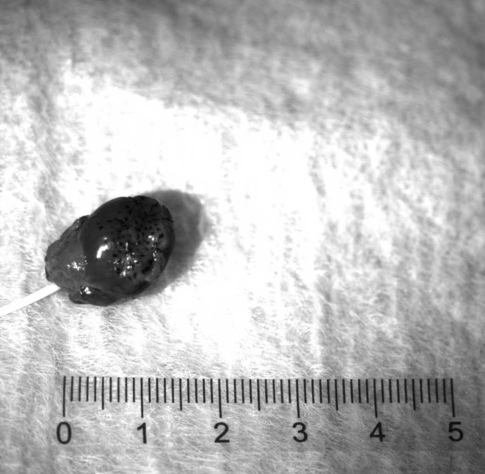
To meet the above requirements, Haytham adopted binocular 3D visual strain gauge to perform non-contact 3D full-field deformation measurement on the cardiac patch.
(1) Test configuration
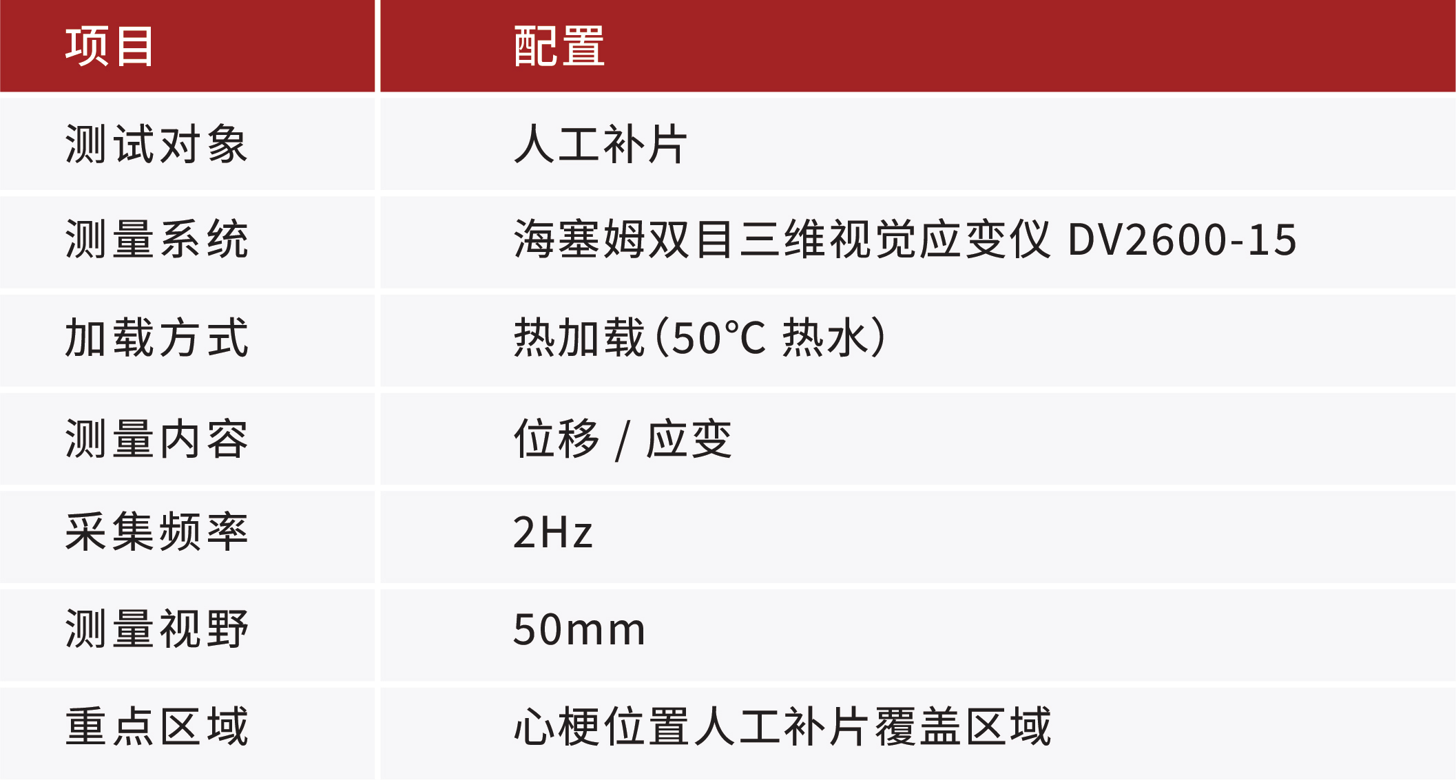
(2) Test preparation
Before the test, the engineers prepared speckles suitable for image correlation calculation on the specimen surface according to the specimen size, measuring range and deformation characteristics, and completed camera arrangement, measurement area setting and system calibration.
During the test, the DIC system continuously captured images of the specimen surface, tracked the position changes of the speckle regions with the digital image correlation algorithm, and computed the displacement and strain information on the specimen surface. The entire process was non-contact, adding no stiffness and leaving the mechanical state of the specimen itself unchanged.
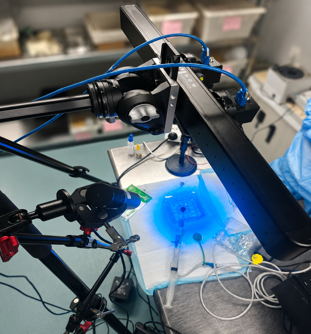
After the test started, the customer continuously applied 50°C hot water to the cardiac patch according to the preset conditions. The binocular DIC synchronously recorded the entire process of the specimen from the initial state to the end of the test, and generated displacement contour maps, strain contour maps and related data curves.
▪ Early loading stage: the temperature had not yet risen to 50°C, the overall deformation of the patch was smooth, and the strain differences among regions were small;
▪ Mid-loading stage: as the temperature gradually rose, obvious displacement began to appear at the myocardial infarction patch position, and local strain gradually concentrated;
▪ Late loading stage: the high-strain region further expanded toward the upper left of the heart, and obvious deformation finally appeared in that region.
The test results show that the cardiac patch exhibited obvious shrinkage deformation after heating.
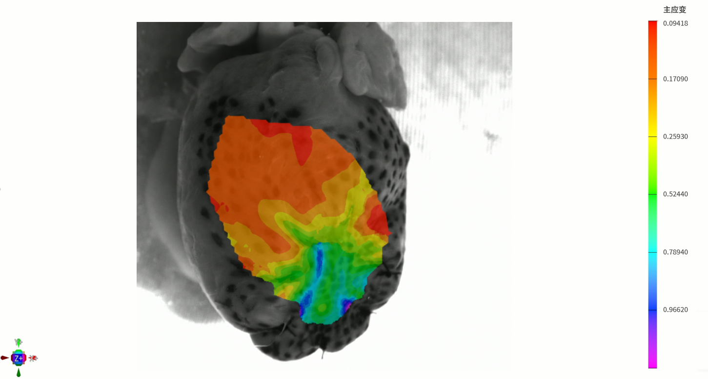
▪ Full-field displacement contour map: the displacement change at the upper left of the myocardial infarction site was the most obvious, and the maximum displacement was concentrated in the upper-left region of the heart;
▪ Strain contour map: strain concentration appeared early at the upper left of the heart, and severe shrinkage occurred as the heating time extended.
Combined with data extraction from the customer's key observation regions, the key results of this test are as follows:
▪ Maximum displacement: 2.174mm
▪ Strain concentration region: upper left of the heart
▪ Key position variation trend: when the temperature reached 50°C, the patch shrank sharply toward the upper left of the heart until reaching the shrinkage limit
These results provided intuitive and quantifiable data for the customer's subsequent material improvement.
In this test, Haytham binocular 3D visual strain gaugedid not merely record that the patch "had deformed", but transformed the entire deformation process into quantifiable, analyzable and traceable displacement and strain data:
▪ Full-field measurement: data over the entire measurement area acquired in a single pass, with no need to guess the high-risk locations in advance;
▪ Non-contact: no bonding, no added stiffness, suitable for flexible, small-sized and wet specimens;
▪ Full-process recording: the deformation evolution from the early loading stage to the shrinkage limit is fully traceable.
As a result, the customer was able to identify local strain concentration and potential weak locations, gaining experimental support for material selection and structural improvement and accumulating reliable scientific data for material R&D in the cardiac surgery industry.


 Downloads

